

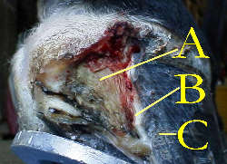
Horse Hoof Anatomy
A = Sensitive Laminae
B = Hoof Horn
C = Hoof Wall
The hoof is layered from the outside in, so the wall of the hoof bears the weight rather than the sole. Like your fingernail, the hoof is made up of dead cells. Dead cells won't heal, so damaged areas must grow out instead of together.
Beneath the hoof wall is the "Hoof Horn" which is attached to the sensitive laminae and attached to the third phalanx, a major bone in the hoof.
At the top of the hoof is the coronary band, the source of nutrition for the hoof wall, similar to the cuticle of the fingernail. The hoof wall consists of tiny moisture retaining tubes of "Keratin" a strong insoluble protein, which grow down from the coronary band at the rate of ¼" to 5/8" per month. The periople is a natural protective coating on the hoof wall, which seals in moisture.
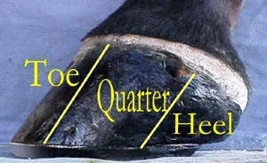
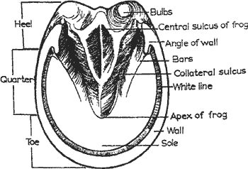
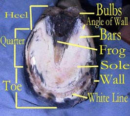
The front side of the hoof is called the toe, the sides are called the quarters and the back of the hoof is called the heel. The hoof wall and hoof horn meets the sole at the bottom of the hoof. The backside of the sole to the middle portion is called the "Frog" a rubbery mass, wedge shaped that acts like a shock absorber. The frog also helps circulate blood within the hoof through a pumping action as the hoof hits the ground.
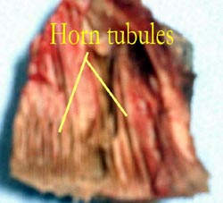
The wall of the hoof grows from the epithelium covering the coronary dermis, which consists of the horn tubules embedded in the intertubular horn that is attached to the coffin bone and hoof cartilage's.
The three basic layers of the structure consist of the stratum externum,meduim and internum. The stratum externum consists of the horn tissue produced by the perioplic dermis, which lies directly proximal to the coronary dermis. The stratum externum is only a few millimeters thick, is somewhat rubbery near the coronary band and is dehydrated over the distal hoof wall.
The bulk of the hoof wall is made up of the stratum medium's pigmented horn tubules. The non- pigmented stratum internum consist of the approximately 600 laminae that interdigitate with the sensitive laminae of the laminar dermis. The dermis of the sole is firmly attached to the undersurface of the coffin bone and produces a mixture of horn tubules and intertubular horn. The junction between the sole and wall is the so - called "white line". The white line includes some of the non-pigmented stratum medium of the wall, the distal ends of the horny laminae and, between these, pigmented horn produced over the terminal papillae of the laminae dermis.